4.4 Language in the Brain
Dinesh Ramoo
The specialization of various parts of the brain would have been a concept you may have come across in other psychology courses. While people have often speculated on these issues in the past, it wasn’t until the 19th century that we really started to gain a scientific understanding of this concept. Now we have even more advanced neuroimaging techniques that have opened new avenues for exploring language. We know that the brain is divided into two hemispheres which are specialized for different functions. In most right-handed people, the left hemisphere is specialized for language processing and production. This is even true of people who used sign-language (Corina et al., 2003).
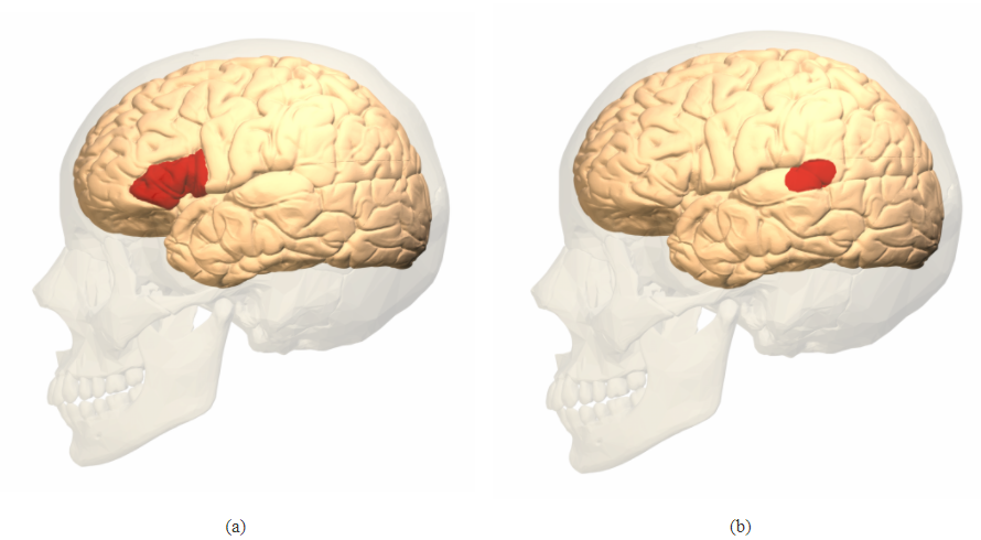
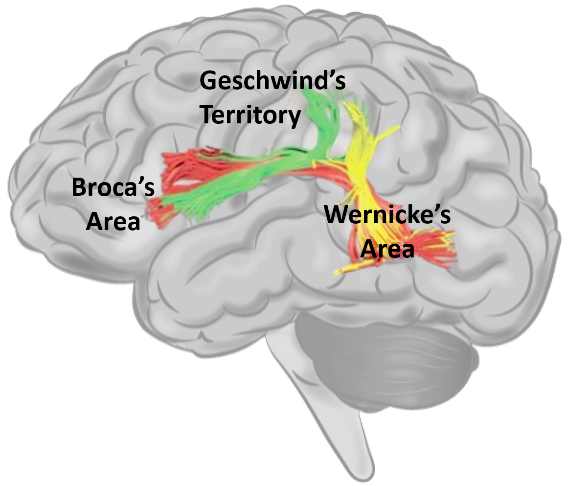
The earliest record of brain damage leading to language loss or aphasia is in an Ancient Egyptian medical texts now known as the Edwin Smith surgical papyrus from 1700 BCE (Minagar et al., 2003). Cases 20 and 22 of the papyrus talk about an individual with an injury on the left side of their skull. He is described as speechless, “an ailment not to be treated.” Such an impairment to language production was not investigated in detail again until Paul Broca explored such issues in the 1860s. Broca observed several patients who had speech disorders resulting from damaged to the left frontal lobe. Later post-mortem examination revealed damaged to an area now known as Broca’s region (see Figure 4.3a). In 1874, a German neurologist Carl Wernicke observed language comprehension issues that arose from damage to the left temporal lobe. The region is know known as Wernicke’s region (see Figure 4.3b). He also proposed the first neurological model of language. As seen in Figure 4.4, we now know that the interpretation of language is associated with Wernicke’s region and it is connected to Broca’s region via a group of nerve fibres known as the arcuate fasciculus. Broca’s region itself is associated with articulation and the controlling of speech. Geschwind (1972) further elaborated on this to describe the Wernicke–Geschwind model. This model brings together what was known about language at the time to describe language production (Broca’s region), comprehension (Wernicke’s region), reading (visual cortex) and spelling (angular gyrus).

This view of the neuroanatomy of language is too simplistic and las its limitations. Language is not always localized to the left hemisphere and the right hemisphere is known to carry out some language functions. Subcortical regions may also have a role to play in language. Even within the cortex, areas other than Broca’s and Wernicke’s region appear to play a role in language processing. With advances in neuroimaging techniques, our understanding of the neuroanatomy of the brain will continue to expand.

Media Attributions
- Figure 4.3 Broca’s Area (a) and Wernicke’s Area (b) by the Database Center for Life Science(DBCLS) is licensed under a CC BY-SA 2.1 Japan licence.
- Figure 4.4 Language Areas of the Brain by Dinesh Ramoo, the author, is an adapted version of Human Brain by Injurymap and Three Segments of the Arcuate Fasciculus by Marco Catani et al., is licensed under a CC BY 4.0 licence.
An acquire disorder which affect language processing after brain trauma.
A region in the frontal lobe of the brain and usually found in the left hemisphere linked to speech production.
A region in the temporal lobe of the brain and usually found in the left hemisphere linked to speech comprehension.

